

November 2014: An incorrect diagnosis sparks the beginning of a nightmare
“Your peripheral vision is black, you have intolerable eye pain, a prolonged headache, an earache and you’re tired? Those are all symptoms of an ear infection called Otitis Media. Here are some ear drops!”
It had now been a week since my first onset of symptoms. I went to work at my local restaurant with poor vision and excruciating pain. After continuing to bump into everything that was in my way and not being able to see, my employer decided to cut my shift short. I entered my car to drive back home and thought that there was a complete blackout in my suburb. I have no idea how I made it home safely but luckily my work was just around the corner. I took a shower, went to sleep and thought to myself that the lights will be back onInflammation of the optic nerve that may be classified as unilateral (affecting one eye) or bilateral (affecting both eyes) that may result in vision changes, vision loss, and/or pain with eye movement. by tomorrow morning. I woke up that morning and everything was black and gray. I took a few steps and fell down the stairs. My parents were immensely worried and ran to help me. At that moment, I realised that there was no blackout in my suburb but a blackout was happening within my vision.
My mum instantly drove me to my GP who turned around and said, “Take her to the Emergency Room now, your daughter has gone blind!”
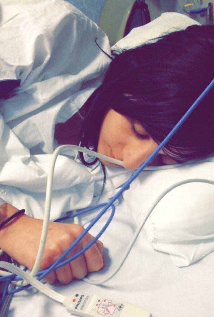
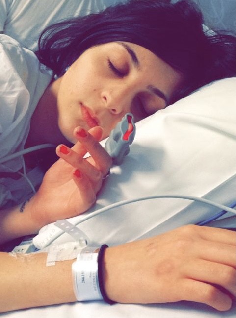
I was admitted to hospital for 5 days and an investigation for my presentation began. An Magnetic Resonance ImagingA noninvasive imaging technique that uses strong magnetic fields to produce images of nearly any structure of the body. For MOGAD, it is typically used with and without contrast to identify disease activity in the central nervous system. (MRIA noninvasive imaging technique that uses strong magnetic fields to produce images of nearly any structure of the body. For MOGAD, it is typically used with and without contrast to identify disease activity in the central nervous system.) at the time revealed that the intra-orbital segments of the optic nervesThe cranial nerves that relay messages from your eyes to your brain to create visual images. These nerves extend from the retina in the back of the eyes to the part of the brain that processes what we see. In myelin oligodendrocyte glycoproten antibody disease (MOGAD), they may be the target of inflammation or lesions, sometimes causing visual disruption or blindness. were swollen bilaterally with signal hyperintensity and enhancement. During the clinical examination, I had bilateralRefers to disease or attack on two sides. optic disc swelling, poor Visual Acuity (VA) bilaterally and I couldn’t see anything onInflammation of the optic nerve that may be classified as unilateral (affecting one eye) or bilateral (affecting both eyes) that may result in vision changes, vision loss, and/or pain with eye movement. the Ishihara chart. My Visual Field Index (VFI) was 27% in the left eye and 4% in the right eye. I was given 3 doses of IntravenousA way of giving a drug or other substance through a needle or tube inserted into a vein. MethylprednisoloneA type of medication typically given intraveneously or orally. For myelin oligodendrocyte glycoprotein antibody disease (MOGAD) it is used as an immunosuppressant and anti-inflammatory to reduce disease activity and inflammation. In adults, it is sometimes used in high doses for acute attacks. In some adult patients, it may be used for 1-2 months after an acute attack to avoid a rapid onset of relapse. Higher doses (>10mg/day) are not recommended for an extended period of time but lower doses (<=10mg/day) may be used in some patients longer term. In children, it may be used after treatment of an acute attack, but it is not recommended long-term due to the profound effects of chronic steroids on a child’s health. Use of oral prednisone as a maintenance therapy in relapsing pediatric MOGAD patients is discouraged due to side effects in developing children, and other therapies such as monthly immune globulin are suggested. (To understand relapsing during steroid tapering, see Steroid Dependency) (IVMP) under the Optic NeuritisInflammation of the optic nerve that may be classified as unilateral (affecting one eye) or bilateral (affecting both eyes) that may result in vision changes, vision loss, and/or pain with eye movement. Treatment Trial (ONTT) protocol.

Diagnosis: Simultaneous BilateralRefers to disease or attack on two sides. Optic NeuritisInflammation of the optic nerve that may be classified as unilateral (affecting one eye) or bilateral (affecting both eyes) that may result in vision changes, vision loss, and/or pain with eye movement. (BON) with suspected autoimmuneA disease in which the immune system incorrectly targets and attacks an individual’s own healthy cells. etiology; a clinically and radiologically isolated presentation.
All my antibody tests were negative and I was given up to 5 years to develop a demyelinating condition known as Multiple Sclerosis (MS). I was a complete mystery. Apart from the recurring intense flashbacks that I couldn’t confirm, I don’t remember much of the admission except what had happened leading up to the admission. My brain decided to click the “delete” button onInflammation of the optic nerve that may be classified as unilateral (affecting one eye) or bilateral (affecting both eyes) that may result in vision changes, vision loss, and/or pain with eye movement. everything associated with that hospital stay. I obtained my medical records for the 2014 presentation in February 2021, that confirmed the presentation, flashbacks and stories associated with the 2014 admission by my parents.
The only positive memory that lingers in my mind from that hospital stay is praying that if I got my vision back, I would become a Primary Teacher.
The next day, my vision returned in my left eye and I immediately enrolled into Australian Catholic University, Strathfield for the Primary Teaching course. My vision returned completely in the right eye just before my first day of University. I had moved onInflammation of the optic nerve that may be classified as unilateral (affecting one eye) or bilateral (affecting both eyes) that may result in vision changes, vision loss, and/or pain with eye movement. with my life and left everything associated with that presentation behind me.
August 2019: Caught in the middle of a tennis match
I was half way through the year as a full-time, Year 1 teacher and my vision in the left eye wasn’t quite right. I had seen my regular Optometrist 2 weeks prior and I was fine, but a decline in my vision was confirmed. A worried Optometrist referred me to a Retinal Specialist as he thought I was having a Retinal Detachment due to the flashing lights and reduced visual acuity. The Retinal Specialist conducted an Optical Coherence Tomography (OCT)A noninvasive imaging technology used to obtain high resolution cross-sectional images of the retina. The layers within the retina can be differentiated and retinal thickness can be measured to aid in the early detection and diagnosis of retinal diseases and conditions such as damage causd by optic neuritis in MOGAD., Humphrey Visual Field (HVF) test and a clinical examination. “I’m sorry Miss Khazen but you don’t have a Retinal Detachment, you have something called Optic Neuritis.” At that very moment, I felt that time had stood still. He sent me to the hospital for Neurology, 3 doses of IVMP, an MRIA noninvasive imaging technique that uses strong magnetic fields to produce images of nearly any structure of the body. For MOGAD, it is typically used with and without contrast to identify disease activity in the central nervous system., bloodwork and a spinal tapA procedure that involves inserting a needle into the spinal canal to collect spinal fluid, typically for diagnostic purposes. Often used during diagnosis of neurological conditions. Otherwise known as a lumbar puncture.. I was admitted for a few days and blood work was taken for everything except the Myelin Oligodendrocyte Glycoprotein (MOG)A type of protein involved in cell adhesion. Present throughout myelin sheaths. antibodyA protective protein produced by your immune system that attaches to antigens (foreign substances), such as bacteria and toxins, and removes them from your body. In myelin oligodendrocyte glycoproten antibody disease (MOGAD), the body incorrectly produces an antibody that targets myelin oligodendrocyte glycoprotein, a component of the myelin sheath in the central nervous system.. Instead, Myelin-Associated Glycoprotein (MAG), Neuromyelitis Optica (NMO) and other antibodyA protective protein produced by your immune system that attaches to antigens (foreign substances), such as bacteria and toxins, and removes them from your body. In myelin oligodendrocyte glycoproten antibody disease (MOGAD), the body incorrectly produces an antibody that targets myelin oligodendrocyte glycoprotein, a component of the myelin sheath in the central nervous system. tests were ordered and results were returning negative. The MRIA noninvasive imaging technique that uses strong magnetic fields to produce images of nearly any structure of the body. For MOGAD, it is typically used with and without contrast to identify disease activity in the central nervous system. revealed slight enhancement of my optic nervesThe cranial nerves that relay messages from your eyes to your brain to create visual images. These nerves extend from the retina in the back of the eyes to the part of the brain that processes what we see. In myelin oligodendrocyte glycoproten antibody disease (MOGAD), they may be the target of inflammation or lesions, sometimes causing visual disruption or blindness. and no brain lesions. Neurology and Ophthalmology couldn’t agree onInflammation of the optic nerve that may be classified as unilateral (affecting one eye) or bilateral (affecting both eyes) that may result in vision changes, vision loss, and/or pain with eye movement. a diagnosis, giving me a spinal tapA procedure that involves inserting a needle into the spinal canal to collect spinal fluid, typically for diagnostic purposes. Often used during diagnosis of neurological conditions. Otherwise known as a lumbar puncture. or treatment.
I felt like a tennis ball caught in the middle of a never ending tennis match between Ophthalmology and Neurology.
I was discharged a few days later with no diagnosis or treatment and was told that I was a difficult patient, not because I was difficult but because they didn’t know what was wrong with my vision. My family members and I in complete disbelief packed everything and left with heightened emotions.
I never thought I would hear the words "Optic Neuritis" ever again
My visual acuity kept declining in the left eye, patterns seemed unusual, colours were dull and lights were dim. I went back to my GP who sent me to an OphthalmologistA board-certified medical doctor that specializes in diseases and disorders of the eye. as I didn’t want to return to the previous hospital after my experience. After conducting the usual eye examinations, I was told that I definitely have Optic NeuritisInflammation of the optic nerve that may be classified as unilateral (affecting one eye) or bilateral (affecting both eyes) that may result in vision changes, vision loss, and/or pain with eye movement. and was sent to another hospital that he currently worked at. They immediately called the Ophthalmologist there and the on-call Neurology team where I was admitted for 3 doses of IVMP, blood work and a spinal tap. They obtained my medical records between 2014-2019 from the previous hospital and began investigating what caused this ONInflammation of the optic nerve that may be classified as unilateral (affecting one eye) or bilateral (affecting both eyes) that may result in vision changes, vision loss, and/or pain with eye movement. relapseWhen you present to your doctor or hospital with new or worsening central nervous system symptoms. Generally, if your symptoms gradually worsen over 24-48 hours, there is heightened concern of a relapse. (Also referred to as a flare by the myelin oligodendrocyte glycoproten antibody disease (MOGAD) community).. At the time, my MOGA type of protein involved in cell adhesion. Present throughout myelin sheaths. antibodyA protective protein produced by your immune system that attaches to antigens (foreign substances), such as bacteria and toxins, and removes them from your body. In myelin oligodendrocyte glycoproten antibody disease (MOGAD), the body incorrectly produces an antibody that targets myelin oligodendrocyte glycoprotein, a component of the myelin sheath in the central nervous system. test had not yet returned. I was given 50 mg of Oral PrednisoneA type of medication typically given intraveneously or orally. For myelin oligodendrocyte glycoprotein antibody disease (MOGAD) it is used as an immunosuppressant and anti-inflammatory to reduce disease activity and inflammation. In adults, it is sometimes used in high doses for acute attacks. In some adult patients, it may be used for 1-2 months after an acute attack to avoid a rapid onset of relapse. Higher doses (>10mg/day) are not recommended for an extended period of time but lower doses (<=10mg/day) may be used in some patients longer term. In children, it may be used after treatment of an acute attack, but it is not recommended long-term due to the profound effects of chronic steroids on a child’s health. Use of oral prednisone as a maintenance therapy in relapsing pediatric MOGAD patients is discouraged due to side effects in developing children, and other therapies such as monthly immune globulin are suggested. (To understand relapsing during steroid tapering, see Steroid Dependency) to slowly taper for 3 months but unfortunately only lasted 1 month onInflammation of the optic nerve that may be classified as unilateral (affecting one eye) or bilateral (affecting both eyes) that may result in vision changes, vision loss, and/or pain with eye movement. them due to the side effects. I was positive for the Bartonella Henselae infection in August and severely symptomatic by September 1, with a fever, an inflamed ear and left-side parotitis, among other symptoms.
I was given antibiotics and a possibility was raised that this could’ve triggered or caused the UnilateralRefers to disease or attack on a single side. Optic NeuritisInflammation of the optic nerve that may be classified as unilateral (affecting one eye) or bilateral (affecting both eyes) that may result in vision changes, vision loss, and/or pain with eye movement. (UON) relapseWhen you present to your doctor or hospital with new or worsening central nervous system symptoms. Generally, if your symptoms gradually worsen over 24-48 hours, there is heightened concern of a relapse. (Also referred to as a flare by the myelin oligodendrocyte glycoproten antibody disease (MOGAD) community)..
My dream finally came true when I graduated University with a Bachelor of Education - (Primary) Degree in 2019


March 2020: Another relapse leads to a diagnosis of Myelin Oligodendrocyte Glycoprotein Antibody - Associated Disease (MOGAD)
I presented to the hospital once again with similar symptoms to those in August 2019, and after many tests I was seated by two Ophthalmologists who handed me a yellow sticky note that said, “Optic Neuritis caused by MOG.” I was positive for the MOGA type of protein involved in cell adhesion. Present throughout myelin sheaths. antibodyA protective protein produced by your immune system that attaches to antigens (foreign substances), such as bacteria and toxins, and removes them from your body. In myelin oligodendrocyte glycoproten antibody disease (MOGAD), the body incorrectly produces an antibody that targets myelin oligodendrocyte glycoprotein, a component of the myelin sheath in the central nervous system. in both serum and Cerebrospinal Fluid (CSF)A clear, colorless liquid found throughout the cavities of the central nervous system that helps protect the brain and spinal cord by acting like a cushion against sudden impact or injury, and provides a medium for the transport of nutrients and the removal of waste products for proper functioning. during the August 2019 presentation onInflammation of the optic nerve that may be classified as unilateral (affecting one eye) or bilateral (affecting both eyes) that may result in vision changes, vision loss, and/or pain with eye movement. a Live cell-based assayA laboratory test used to: • Measure the presence or amount of a specific substance: in a sample, such as blood, urine, or tissue.
• Determine the potency or activity: of a drug or other substance.
• Analyze the purity: of a chemical compound.
• Identify and quantify: biomarkers related to a disease or condition.
and I was receiving the diagnosis 7 months later! The Ophthalmologists kept persisting that there was no such thing as BilateralRefers to disease or attack on two sides. Optic NeuritisInflammation of the optic nerve that may be classified as unilateral (affecting one eye) or bilateral (affecting both eyes) that may result in vision changes, vision loss, and/or pain with eye movement. (BON) and that they knew nothing about MOGA type of protein involved in cell adhesion. Present throughout myelin sheaths. as all my symptoms were too severe and atypical for ONInflammation of the optic nerve that may be classified as unilateral (affecting one eye) or bilateral (affecting both eyes) that may result in vision changes, vision loss, and/or pain with eye movement.. After consulting with the on-call Neurologist, he ordered 3 doses of IVMP and referred me to a Neurologist as this hospital did not have a Neurology Clinic. The hospital rotates different Ophthalmologists every 6 months – 1 year, so they never have the same doctors. A lot of people would’ve celebrated after finally having a diagnosis, but I was confused, deflated and had many questions. No such thing as BON? Symptoms too severe and atypical for ONInflammation of the optic nerve that may be classified as unilateral (affecting one eye) or bilateral (affecting both eyes) that may result in vision changes, vision loss, and/or pain with eye movement.? A doctor didn’t know what MOGA type of protein involved in cell adhesion. Present throughout myelin sheaths. was? I was then referred to Concord Hospital after another Neurologist wasn’t too familiar with MOGADOften referred to as MOGAD, Anti-MOG, MOG Ab+, MOG Antibody Disease, MOG Associated Antibody Disease, MOG positive disease .
Family is forever: The ones who truly matter
Throughout my difficult journey, I’ve been so thankful for my parents, brothers, extended family members and friends who’ve been an amazing support system. They’ve been by my side through the hospital stays, tears, frustration, laughter, graduation and without them I wouldn’t be the woman that I am today. My mum, cousin Debie and aunty Mirna have been there through it all. I’m so appreciative for these 3 inspirational women in my life as I wouldn’t have been able to do any of this without them.
Words cannot express how much I love them all so endlessly as they have been the glue holding me together throughout those challenging years.


My incredible medical team - Two shining lights instead of one at the end of the tunnel
I met Dr. Sudarshini Ramanathan (Darshi) or as I call her Dr. Darsh, and Associate Professor Sean Riminton at Concord Hospital in May, 2020. Having experienced such a diabolical journey with the medical system and a rollercoaster of emotions, my walls were up and cemented with bricks. I knew nothing about MOGAD and my confidence was down from the previous comments made by the 2 Ophthalmologists in regards to my presentations and the disease itself. But Dr. Darsh and Associate Professor Sean reassured me that my diagnosis was consistent with MOGAD clinically, radiologically and a positive serum and CSFA clear, colorless liquid found throughout the cavities of the central nervous system that helps protect the brain and spinal cord by acting like a cushion against sudden impact or injury, and provides a medium for the transport of nutrients and the removal of waste products for proper functioning. test onInflammation of the optic nerve that may be classified as unilateral (affecting one eye) or bilateral (affecting both eyes) that may result in vision changes, vision loss, and/or pain with eye movement. a Live cell-based assayA laboratory test used to: • Measure the presence or amount of a specific substance: in a sample, such as blood, urine, or tissue.
• Determine the potency or activity: of a drug or other substance.
• Analyze the purity: of a chemical compound.
• Identify and quantify: biomarkers related to a disease or condition.
was the icing onInflammation of the optic nerve that may be classified as unilateral (affecting one eye) or bilateral (affecting both eyes) that may result in vision changes, vision loss, and/or pain with eye movement. the cake confirming the diagnosis. I knew that my walls weren’t going to come down that easily as I was traumatised and a sense of frustration and mistrust was brewing inside of me from my previous experiences with the medical system, so I began not taking my diagnosis seriously. However, Dr. Darsh and Associate Professor Sean did not give up onInflammation of the optic nerve that may be classified as unilateral (affecting one eye) or bilateral (affecting both eyes) that may result in vision changes, vision loss, and/or pain with eye movement. me, especially when I grappled to come to terms with my diagnosis. Instead, they were patient, understanding and helped me take small but valuable steps towards my diagnosis.
Between April – June 2021, Dr. Darsh communicated 4 important things to me:
1. Knowledge is power and WE are in a position of strength because WE have a diagnosis.
2. These are your hand of cards and it’s what WE do with them that truly matters.
3. WE are a team who want the best outcome for you and WE want to make sure that you are staying well.
4. When you are having symptoms, please come or let me know so WE can work on this together.
Everytime I heard the word “WE” I knew that I was not in this by myself anymore. Dr. Darshi and Associate Professor Sean have been absolutely amazing. Dr. Darshi has been an advocate for me and has also taught me how to advocate for myself. Our journey has consisted of a lot of laughter, tears, relapsesWhen you present to your doctor or hospital with new or worsening central nervous system symptoms. Generally, if your symptoms gradually worsen over 24-48 hours, there is heightened concern of a relapse. (Also referred to as a flare by the myelin oligodendrocyte glycoproten antibody disease (MOGAD) community)., flare ups, trying to find the right treatment plan, hospital visits and my highly cemented brick walls. But I can whole-heartedly say that I’m in good hands and feel confident in their care. I have so much love and respect for the both of them and could not have asked for a better team of specialists.
Between the ages 2-5, I had also experienced a complex medical journey at such a young age in Lebanon before moving to Australia in December 1999. I suffered from infections and fevers that led to hospitalisations with visual issues, balance problems, irritability and more. Due to the lack of medical tools and specialist knowledge back then, I was unable to be given a diagnosis but my current specialists feel that I could’ve potentially had Acute Disseminated Encephalomyelitis (ADEM)An initial and brief autoimmune attack that causes widespread inflammation and damage to the brain, spinal cord, and optic nerve, typically in children. This initial attack establishes the basis for diagnosis. The MOG antibody is detectable in a percentage of Acute Disseminated Encephalomyelitis (ADEM) patients..
The MOG Project - A blessing in disguise
Where do I start with this wonderful group of people especially Julia, Peter and Andrea? When I met them I was confused, full of questions and struggling to come to terms with my diagnosis. It’s incredible how a group of people can come together internationally with the same disease. I began to ask questions, educate myself by reading a lot of research articles, listening to podcasts and viewing webinars about MOGADOften referred to as MOGAD, Anti-MOG, MOG Ab+, MOG Antibody Disease, MOG Associated Antibody Disease, MOG positive disease . I was on a mission to ensure that no child or adult ever had to feel the same way that I did throughout my journey towards a diagnosis. A relationship with them was formed when I put forward a plan to raise awareness for MOGADOften referred to as MOGAD, Anti-MOG, MOG Ab+, MOG Antibody Disease, MOG Associated Antibody Disease, MOG positive disease from an Educator’s perspective but also when they found out I was quite hilarious. I’m honoured to be a Lebanese-Australian Primary Teacher onInflammation of the optic nerve that may be classified as unilateral (affecting one eye) or bilateral (affecting both eyes) that may result in vision changes, vision loss, and/or pain with eye movement. The MOGA type of protein involved in cell adhesion. Present throughout myelin sheaths. Project’s Advisory board and use my expertise in Mathematics to organise and analyse data, my research skills and creativity to raise awareness for MOGADOften referred to as MOGAD, Anti-MOG, MOG Ab+, MOG Antibody Disease, MOG Associated Antibody Disease, MOG positive disease , formulate projects with doctors, The MOGA type of protein involved in cell adhesion. Present throughout myelin sheaths. Project members and partners and expand social media networks.
I have nothing but love for this bunch and without a doubt, they’ve become my second international family.
It’s time for me to play the right hand of cards that will help me win against this rare immune-mediated neurological disorder called MOGAD!
My journey has been difficult, far from a straight road and at times, a lot to take onInflammation of the optic nerve that may be classified as unilateral (affecting one eye) or bilateral (affecting both eyes) that may result in vision changes, vision loss, and/or pain with eye movement.. If I hadn’t lost my vision in 2014 then regained it, I definitely wouldn’t be the Primary Teacher that I am today, who’s an advocate for little people, passionate about my career and loves educating young children. I strongly believe that there are two options in life. When life hands you a set of cards, you need to decide whether you want to keep folding or finally play the game and see where it gets you!
I have finally entered the MOGADOften referred to as MOGAD, Anti-MOG, MOG Ab+, MOG Antibody Disease, MOG Associated Antibody Disease, MOG positive disease game with a set of cards that consists of faith, an amazing family, a great support system from The MOG Project, treatment plans and an incredible, kind, caring, patient and highly educated Neurologist and ImmunologistA doctor or scientist who specializes in immune system disorders..
It’s time for me to win!

By Jenny Khazen


Hi Jenny,
A family member was recently diagnosed with MOG, I would appreciate further conversation with you. Your article was very interesting and I would love to learn more about project MOG.
Thank you!
Comments are closed.